In embryos, discrete organs and entire body plans emerge from the coordinate actions of individual cells. Whether these cells comprise highly organized epithelia or more loosely associated mesenchyme, the execution of major morphogenetic events requires dynamic regulation of cell shape, polarity and adhesion across cell populations. We are interested in how these diverse cellular behaviors are orchestrated to generate form. Our lab employs genetic, cell biological and biochemical techniques to elucidate the molecular mechanisms that sculpt tissues and organs during development.

Drosophila egg chamber development as a model for organ morphogenesis

We are currently investigating the morphogenetic mechanisms that create the elliptical shape of the Drosophila egg. Eggs form within the ovaries of adult female flies from multicellular units known as egg chambers. The basic structure of an egg chamber is quite simple. There is an internal germ cell cluster, consisting of a single oocyte and 15 nurse cells, which is surrounded by an epithelium of ~650 follicle cells. Because the oocyte is the only cell present in the mature egg, the primary function of the other cell types is to pattern and nurture this one cell. During this process, however, the egg chamber undergoes dramatic morphogenesis that rivals the formation of any complex organ in the embryo.

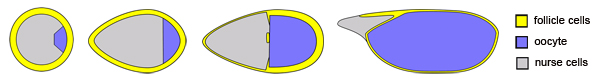
Changes in egg chamber morphology over time (images not drawn to scale)

One prominent morphogenetic change involves the lengthening of this organ along its anterior-posterior axis, which transforms the initially spherical egg chamber into an elliptically shaped egg. Through a forward genetic screen, we have identified a large collection of mutations that disrupt this elongation process, leading to the production of spherical eggs. This collection of ‘round egg’ mutants provides an unprecedented opportunity to gain molecular insight into this poorly understood morphogenetic process. The two main questions the lab is addressing are detailed below.

How is planar polarity established in the follicle cell epithelium?

Other labs have shown that planar polarity within the FC epithelium is required for egg chamber elongation. This planar polarity takes the form of parallel arrays of actin filaments and extracellular matrix fibrils that encircle the outside of the egg chamber, perpendicular to the anterior-posterior axis. Interestingly, FC planar polarity does not depend on the Frizzled-PCP signaling pathway, suggesting that an unconventional regulatory mechanism operates in this tissue. We wish to understand the molecular underpinnings of this novel planar polarity system and are particularly interested in testing the hypothesis that the polar cells, a highly specialized cell type in the egg chamber, induce planar polarity in the neighboring FCs.

What are the cellular behaviors that drive egg chamber elongation?

Once planar polarity is established in the FCs, this positional information is used to coordinate the individual cellular behaviors that ultimately drive egg chamber elongation. Very little is known, however, about what these polarized cellular behaviors might be. Well known mechanisms for tissue elongation including: directed cell shape change, polarized cell division, and cell intercalation, do not occur in the FCs during the elongation process, suggesting that another cellular mechanism controls this morphogenesis. We are using newly developed live imaging techniques, in both wild-type and mutant contexts, to better understand the polarized FC behaviors that drive elongation morphogenesis in the egg chamber.

Extension of principles discovered in Drosophila to vertebrates

The ultimate goal of this research is to understand general principles governing organ morphogenesis and how the impairment of these functions contributes to human disease. The unrivaled genetic manipulability of Drosophila makes it a premier system for gene discovery and sophisticated dissection of molecular mechanisms. However, to understand the human relevance of discoveries made in Drosophila requires translation to a vertebrate system. Zebrafish are highly amenable to reverse genetics and live imaging, making them the ideal vertebrate complement to forward genetic studies of morphogenesis in flies. As the lab matures, we will use reverse genetics in zebrafish as a gateway to investigate the vertebrate functions of the morphogenetic regulators we identify in Drosophila. Novel insights from the zebrafish work can then feedback into the fly research, achieving a highly synergistic approach to the study of tissue and organ morphogenesis.

Suggested Reading

For additional background on egg chamber elongation, please see the following articles:

Horne-Badovinac, S. & Bilder, D., Mass transit: epithelial morphogenesis in the Drosophila egg chamber. Dev Dyn 232, 559-74 (2005).

Gutzeit, H.O. The microfilament pattern in the somatic follicle cells of mid-vitellogenic ovarian follicles of Drosophila. Eur J Cell Biol 53, 349-56 (1990).

Gutzeit, H.O., Eberhardt, W. & Gratwohl, E. Laminin and basement membrane-associated microfilaments in wild-type and mutant Drosophila ovarian follicles. J Cell Sci 100 (Pt 4), 781-8 (1991).

Bateman, J., Reddy, R.S., Saito, H. & Van Vactor, D. The receptor tyrosine phosphatase Dlar and integrins organize actin filaments in the Drosophila follicular epithelium. Current Biol 11, 1317-27 (2001).

Frydman, H.M. & Spradling, A.C. The receptor-like tyrosine phosphatase lar is required for epithelial planar polarity and for axis determination within drosophila ovarian follicles. Development 128, 3209-20 (2001).